
A new type of 3D printer may be capable of making muscle, bone and other types of tissue that are good enough for implanting in humans, scientists report.
So-called 3D “bioprinters” are machines that can print out cells in layered patterns, with the goal of creating body tissue or even complex organs. But until now, a major stumbling block has been the scale of the printed structures.
“If you try to make something that’s larger, it turns gooey and falls apart,” explained Dr. Glenn Green, an associate professor of pediatric otolaryngology at the University of Michigan.
Another limitation, Green said, has been the lack of blood vessels in bioprinted tissue: Larger structures are not possible without blood vessels to supply nutrients and oxygen.
The new technology, described in the Feb. 15 online edition of Nature Biotechnology, seems to surmount those challenges.
“This is exciting,” said Green, who was not involved in the research, but has studied 3D bioprinting. “This is a big breakthrough in identifying a way to make tissue that is larger and could be applied to humans.”
Ultimately, the hope is to have 3D printers that can churn out any kind of human tissue — to replace tissue damaged by trauma, disease or birth defects, said Dr. Anthony Atala, the senior researcher on the new study.
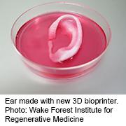
To make that replacement tissue, cells would be taken from a patient’s own body, which should avoid the risk of an immune system attack, said Atala. He directs the Wake Forest Institute for Regenerative Medicine, in Winston-Salem, N.C.
But that’s the goal for the future, and many hurdles remain before the technique might be used in humans.
For now, Atala said, his team has shown that it’s feasible to create ear, bone and muscle tissue that are “human-scale.”
The researchers developed the new printing system over 10 years. It creates a biodegradable, plastic-like material that gives the printed tissue its shape, along with cells suspended in a water-based “ink.” The tissue also has a system of “micro-channels” that allows nutrients and oxygen from the body to diffuse into the structure until a system of blood vessels can form.
Atala’s team found that when it implanted bioprinted bone, muscle and cartilage into rodents, the structures matured into functional tissue, complete with a network of blood vessels.
The researchers also printed a human jawbone fragment that was the right size and shape to be used in facial reconstruction surgery.
According to Green, there’s “no technical barrier” to implanting such printed tissues into humans — but there are some critical questions.
“We don’t know what happens with these tissues long-term,” Green said. Plus, he added, the experiments described in this study used materials that are not approved for use in humans. The jawbone fragment, for instance, was created with stem cells from human amniotic fluid.
As 3D bioprinting moves forward, Green said, it will probably focus first on simpler structures that don’t move or bear weight — including the ears, nose or bones in the skull — before trying to tackle more complicated tissue, or organs such as the heart, kidneys and pancreas.
Atala said his team plans to implant bioprinted cartilage, bone and muscle tissue into patients in the future, with funding from the Armed Forces Institute of Regenerative Medicine. The institute, which partly financed the current study, focuses on using regenerative medicine to treat battlefield injuries.
More information
The U.S. National Institutes of Health has more on regenerative medicine.
Source: HealthDay
Copyright © 2026 HealthDay. All rights reserved.
Leave a Reply